

In real hospital environments, medical imaging is one of the most powerful tools doctors rely on, but it is also one of the most pressure-heavy. Every day, radiologists go through hundreds of CT scans, MRIs, and X-rays, often under time constraints and high patient loads. Even the most experienced specialists can miss subtle signs when fatigue sets in or when cases look almost identical.
The truth is, diagnostic errors in imaging are not rare. They usually happen because early-stage diseases can be extremely subtle. A small lung nodule, a tiny brain lesion, or early signs of a blocked artery can easily blend into background noise, especially in large volumes of scans. This is where accuracy becomes critical. A small improvement in detection can completely change a patient’s outcome.
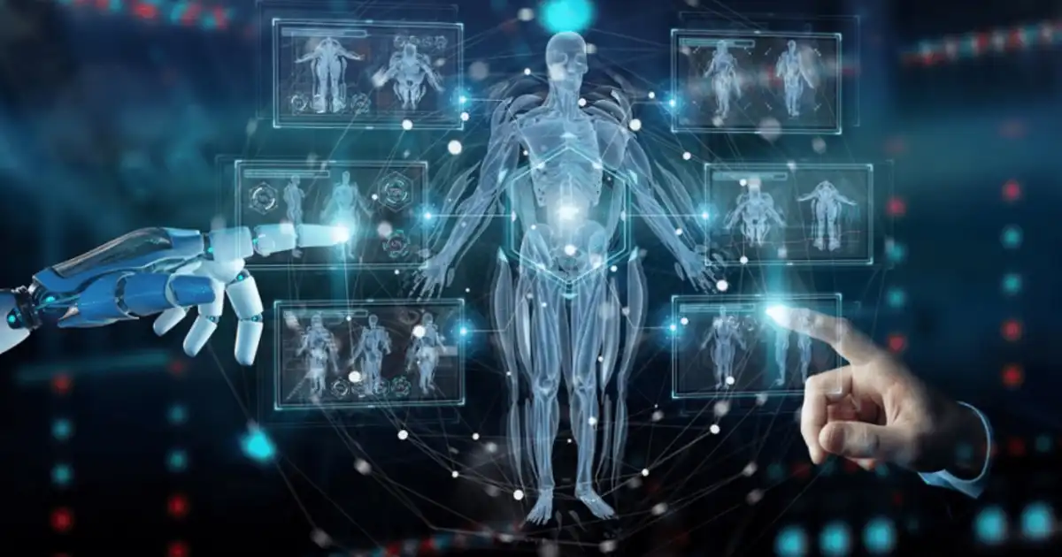
This is exactly where AI medical imaging analysis is starting to make a real difference. In practical hospital settings, AI is not replacing radiologists. Instead, it is being used as a support layer that quietly reviews scans, highlights suspicious regions, and helps doctors double-check their findings.
What I’ve seen in real-world imaging workflows is that AI works best when it acts like a second pair of eyes. It doesn’t get tired, it doesn’t rush, and it can scan thousands of patterns in seconds. But it also depends heavily on data quality and proper clinical integration.
In this article, I’ll break down how AI actually works inside medical imaging systems, where it genuinely improves diagnostic accuracy, and where it still struggles in real clinical environments.
What Is AI Medical Imaging Analysis?
AI medical imaging analysis is basically a system that helps doctors interpret medical images like X-rays, CT scans, MRIs, and ultrasound images using trained algorithms. But in real terms, it is not some magical machine making decisions. It is more like a highly trained assistant that has “seen” millions of past images and learned patterns from them.
In hospitals, these systems are usually integrated into radiology software. When a scan is taken, the image doesn’t just go to the radiologist anymore. It often passes through an AI layer first. That layer scans for abnormalities, measures structures, and flags anything that looks suspicious.
Most of these systems are powered by deep learning in medical imaging, especially neural networks like CNN in healthcare applications. These models are trained on large datasets of labeled images, where experts have already marked tumors, fractures, or other conditions. Over time, the system learns what a normal lung looks like, what a cancerous lesion might resemble, and how subtle changes appear in early disease stages.
But what’s important to understand is that AI does not “understand” images the way humans do. It recognizes patterns. For example, it might detect that a cluster of pixels in a lung scan matches patterns commonly associated with early-stage pneumonia or nodules.
In practice, radiology AI systems are used for three main tasks. First, detection, where the system finds possible abnormalities. Second, segmentation, where it outlines the exact area of concern. Third, classification, where it suggests what the issue might be.
From what I’ve observed, the real value is not in replacing human judgment but in reducing the chance that something important gets overlooked. Especially in busy hospitals, where hundreds of scans come in daily, this extra layer of analysis makes a noticeable difference in workflow efficiency and diagnostic consistency.
How AI Works in Medical Imaging Systems
To understand how AI medical imaging analysis works in real hospital settings, it helps to follow the actual workflow from scan to diagnosis.
First, a patient undergoes imaging, such as an MRI or CT scan. The machine generates raw image data, which is then converted into a digital format stored in systems like PACS (Picture Archiving and Communication System). This is where AI typically enters the pipeline.
Once the image is available, the AI model processes it in several steps. It starts by normalizing the image, which means adjusting brightness, contrast, and scale so that the system can analyze it consistently. Different machines and hospitals produce slightly different image outputs, so this step is essential.
Next comes feature extraction. This is where deep learning in medical imaging really shows its strength. A CNN in healthcare applications breaks the image into layers of patterns. Early layers detect simple features like edges and shapes, while deeper layers identify complex structures like organ boundaries or abnormal tissue formations.
After that, the model runs classification and detection tasks. For example, in a chest X-ray, it may scan for signs of pneumonia, lung nodules, or fluid buildup. It assigns probability scores to each finding. Something might be labeled as “suspicious nodule, 87% confidence.”
Then comes localization, where the system highlights the exact area on the image. This is crucial because doctors don’t just need a label, they need to know where the issue is.
Finally, the output is sent back into the radiologist’s workstation. In many systems I’ve seen, the AI findings appear as overlays on the original scan. The doctor can toggle them on or off, compare interpretations, and decide whether to accept or ignore the AI suggestion.
One important real-world detail is that AI systems are usually trained on specific types of data. A model trained on adult chest X-rays may not perform as well on pediatric scans or rare conditions. That’s why hospitals still validate AI outputs instead of trusting them blindly.
So in practice, AI is not “reading” the scan alone. It is embedded inside a structured workflow that supports the radiologist, not replaces them. The accuracy comes from this combination of machine pattern recognition and human clinical judgment working together.
How AI Improves Diagnostic Accuracy
The biggest claim around AI medical imaging analysis is improved accuracy, but in real hospital environments, this improvement comes from several very practical mechanisms rather than one dramatic breakthrough.
One of the most important improvements is error reduction due to fatigue. Radiologists often work through long queues of scans. After hours of continuous review, even highly trained specialists can miss subtle abnormalities. AI does not suffer from fatigue. It consistently analyzes every image with the same level of attention, whether it is the first scan of the day or the hundredth.
Another major improvement is early-stage detection. Many diseases start with extremely small changes that are hard to see with the human eye. For example, early lung cancer may appear as a faint shadow or tiny nodule. AI systems trained with deep learning in medical imaging can detect these micro-patterns by comparing them against millions of similar cases. In real practice, this often leads to earlier flagging of potential issues, which can significantly improve treatment outcomes.
AI also improves accuracy through image enhancement and preprocessing. In some cases, scans are noisy or unclear due to patient movement or technical limitations. Computer vision in medicine techniques can help enhance contrast, reduce noise, and make hidden details more visible. This does not replace the original scan but gives doctors a clearer version to interpret.
Another practical benefit is the “second opinion” effect. In many hospitals, AI acts like a silent reviewer. After the radiologist completes their assessment, the system cross-checks the findings. If there is a mismatch, it highlights it for review. I’ve seen cases where AI flagged something that was initially missed, prompting a re-evaluation that changed the diagnosis. This doesn’t mean AI is always right, but it adds a safety net.
AI is also extremely good at pattern recognition across large datasets. Humans are good at understanding context, but AI can scan thousands of previous cases and identify statistical similarities that are not obvious. For example, subtle texture changes in tissue that correlate with specific diseases can be detected more consistently by algorithms trained on large datasets.
However, accuracy improvement is not just about detection. It is also about consistency. Two radiologists might interpret borderline cases slightly differently. AI provides a standardized reference point, which helps reduce variability in reporting.
That said, it is important to be realistic. AI can also produce false positives, where normal structures are flagged as abnormal. This is why radiologists always review AI suggestions rather than accepting them automatically.
In real-world clinical use, the best accuracy improvements come from collaboration. AI handles repetitive scanning and pattern highlighting, while doctors apply clinical judgment, patient history, and contextual reasoning. This combination is what actually improves diagnostic reliability, not AI alone.
Real-World Applications
AI medical imaging analysis is already deeply embedded in several medical specialties, and its role varies depending on the type of imaging and disease being studied.
In oncology, AI is widely used for tumor detection and monitoring. It helps identify small growths in organs like the lungs, liver, and breast. Radiology AI systems can also track how tumors change over time, which is crucial for evaluating treatment response. In breast cancer screening, for example, AI assists in mammography interpretation by highlighting suspicious calcifications.
In neurology, AI plays a major role in analyzing brain scans. It can detect signs of stroke, brain hemorrhage, or early neurodegenerative diseases. Time is critical in stroke cases, and AI helps speed up detection so treatment can begin faster. It can also segment brain structures to support research and surgical planning.
In cardiology, AI is used to analyze cardiac CT and MRI scans. It helps measure heart function, detect blocked arteries, and assess structural abnormalities. In some systems, AI can quickly flag high-risk coronary conditions that require urgent attention.
In pulmonology, AI is heavily used for chest X-rays and CT scans. It detects pneumonia, tuberculosis, lung nodules, and even early signs of chronic lung disease. During high patient loads, especially in emergency settings, this support becomes extremely valuable.
In orthopedics, AI helps identify fractures, joint degeneration, and bone misalignments. It is especially useful in emergency rooms where quick triage decisions are needed. AI can quickly highlight possible fractures that may not be obvious in initial scans.
Across all these fields, the core idea remains the same. AI assists by identifying patterns, highlighting abnormalities, and reducing the chances of missed findings.
Benefits
From a hospital workflow perspective, the benefits of AI medical imaging analysis are very practical rather than abstract.
One of the biggest advantages is speed. Radiologists can review AI-processed scans faster because the system already highlights potential issues. This is especially important in emergency departments where time-sensitive decisions are critical.
Another major benefit is improved consistency. Human interpretation can vary depending on experience, workload, and fatigue. AI provides a stable reference point that helps standardize reporting across different doctors and departments.
AI also reduces the cognitive load on radiologists. Instead of scanning every pixel manually, doctors can focus on areas that the system flags. This makes the workflow more efficient and reduces mental exhaustion during long shifts.
In many hospitals, AI also helps prioritize urgent cases. For example, if a scan shows signs of a possible brain bleed or severe lung issue, the system can push that case to the top of the review queue. This triage function is extremely valuable in busy clinical environments.
Another benefit is improved early detection rates. Subtle abnormalities that might be missed in routine review are more likely to be flagged when AI is used as a second layer of analysis.
Finally, AI supports training and education. Junior radiologists can compare their interpretations with AI outputs and senior reviews, helping them learn faster and improve diagnostic skills over time.
Limitations and Challenges
Despite its strengths, AI medical imaging analysis has real limitations that show up in day-to-day hospital use.
One major issue is data bias. If an AI model is trained mostly on certain populations or imaging equipment, it may not perform equally well in other settings. This can lead to inconsistent accuracy across different hospitals or patient groups.
Privacy is another serious concern. Medical imaging data is highly sensitive, and using it to train or run AI systems requires strict compliance with healthcare regulations. Any breach or mishandling of data can have serious consequences.
There are also regulatory delays. Medical AI tools must go through rigorous approval processes before they can be used clinically. This slows down deployment, even when the technology is technically ready.
System dependency is another challenge. If hospitals rely too heavily on AI, there is a risk that clinicians may become overly dependent on it. This can be dangerous if the system fails or produces incorrect outputs.
Real-world failure cases do exist. AI can misinterpret unusual anatomy, rare diseases, or poor-quality scans. In some cases, false positives can lead to unnecessary anxiety or additional testing.
Because of these limitations, hospitals treat AI as a support tool rather than a decision-maker. It is helpful, but not infallible.
AI vs Radiologist Collaboration
In real clinical environments, AI does not replace radiologists. Instead, it changes how they work.
What actually happens is a layered workflow. The AI system first analyzes the scan and highlights potential areas of concern. Then the radiologist reviews both the original image and the AI suggestions.
The doctor still makes the final decision. They consider the patient’s medical history, symptoms, lab results, and clinical context, which AI does not fully understand. This is where human judgment remains essential.
From what I’ve seen in practice, the best results come when radiologists treat AI like a junior assistant. It is helpful for spotting details quickly, but it still needs supervision and verification.
In some cases, AI improves confidence in diagnosis. In others, it raises questions that prompt a second look. Either way, it becomes part of the decision-making loop rather than a replacement.
So the real model is not AI versus radiologist. It is AI with radiologist.
Future of AI Medical Imaging
The future of AI medical imaging analysis is heading toward deeper integration rather than standalone tools.
One clear trend is real-time analysis. Instead of reviewing scans after they are taken, AI will increasingly provide feedback during the imaging process itself, helping technicians adjust parameters immediately.
Another direction is multimodal AI, where imaging data is combined with lab reports, genetics, and patient history. This will make AI-assisted diagnosis more context-aware and clinically useful.
We are also moving toward more explainable AI systems. Right now, many models act like black boxes. Future systems will need to show why they made a certain prediction, especially in healthcare where transparency is critical.
At the same time, integration into hospital workflows will become smoother. AI will be less of a separate tool and more of a built-in layer inside radiology software.
However, adoption will still be gradual. Healthcare is careful by nature, and rightly so. Accuracy, safety, and regulation will continue to shape how fast these systems evolve.
You Might Be Interested In
- What Are Ai-powered Social Welfare Fraud Tools?
- What Is A Feature In Machine Learning?
- How To Erase Objects From Photos With Ai?
- How Does Ai Infrastructure Learning Path Work?
- How Do Ai Systems Learn From Data Over Time?
Conclusion
AI medical imaging analysis is not a futuristic concept anymore. It is already quietly working inside hospitals, supporting radiologists and improving how scans are interpreted. What makes it valuable is not just speed, but its ability to consistently highlight details that might otherwise be missed in busy clinical environments.
At the same time, it is important to stay realistic. AI is not perfect, and it does not understand patients the way doctors do. It works on patterns, probabilities, and data, which means it can make mistakes, especially in rare or complex cases. That is why human oversight remains central to the entire process.
In real-world practice, the most effective use of this technology is collaboration. AI handles repetitive scanning and pattern detection, while radiologists apply clinical judgment and context. Together, they create a more reliable diagnostic process than either could achieve alone.
As the technology continues to evolve, its role will likely expand further into early detection, workflow optimization, and multi-source clinical analysis. But the core idea will remain the same: supporting doctors, not replacing them.
FAQs
How does AI medical imaging analysis actually help doctors in real hospitals?
In real hospital workflows, AI helps by reviewing medical scans like CTs, MRIs, and X-rays and highlighting areas that might need closer attention. It acts as a second layer of review, especially useful when radiologists are handling a large number of cases in a short time.
The key benefit is not replacing doctors but reducing the chance of human oversight. AI can quickly scan images for patterns it has learned from large datasets, which helps doctors focus on the most relevant parts of a scan.
Is AI more accurate than human radiologists?
AI can be extremely accurate in specific tasks, especially when trained on large and high-quality datasets. However, in real clinical practice, it is not universally more accurate than radiologists.
Doctors bring context, patient history, and clinical reasoning, which AI does not fully understand. That is why the best results come from combining both rather than comparing them directly.
What types of diseases can AI detect in medical imaging?
AI is commonly used to detect conditions like tumors, lung infections, fractures, strokes, and heart abnormalities. It is especially effective in identifying patterns in oncology, neurology, cardiology, pulmonology, and orthopedics.
Its strength lies in spotting subtle changes in images that may indicate early stages of disease, which can be harder to detect with the naked eye.
Can AI make mistakes in medical imaging analysis?
Yes, AI can make mistakes. It may produce false positives, where normal tissue is flagged as abnormal, or false negatives, where actual issues are missed. This often depends on the quality of training data and the complexity of the case.
That is why AI is always used as a support tool rather than a final decision-maker in medical imaging workflows.
Will AI replace radiologists in the future?
No, AI is not expected to replace radiologists. Instead, it will continue to assist them by handling repetitive analysis and highlighting potential issues in scans.
Radiologists are still essential because they interpret results in the context of patient history, symptoms, and broader clinical information, which AI cannot fully replicate.
